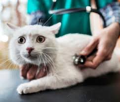
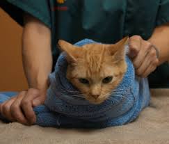
When a patient is fearful of seeing me at a veterinary hospital, their world shrinks to two options: flee or fight. Neither one is fun for them or helps me do what I’m supposed to do for the patient and the family who loves them: find out what’s wrong and make it better.
With patients who are driven by fear, their natural instincts kick in and they resist everything we need to do to get them well.
I’m sure you’ve all been there when trying to treat a fearful patient. Patients displaying aggression are driven by fear. The vast majority of them don’t display this type of behavior at home – so why do they revert to aggression when we are trying to help them?
Think of their first few visits to the hospital and try to see it from their perspective. The car ride. They are likely apprehensive when loaded into the car or carrier. Their owner may try to soothe them with calming words, but fear takes hold.
The sights and smells. When they arrive at the hospital, they can smell the fear from dozens – hundreds – of other pets before them. They can smell the odors of the hospital – cleaning and disinfecting products, the cautery unit in surgery, the parvo dog in isolation. Their heightened sense of smell, many thousands of times what ours is, wafts all of these to them: smells of sickness and death. They sense that this is a bad place.
After this – needles, restraint. The inability to move. The fear of being atop the exam table with no place to run.
Is it any surprise, then, that they would grow to dislike this place and its occupants?
We know that we want to help them. We know that a vaccine will prevent deadly diseases, that a spay will prevent unwanted pregnancies and pyometra…but they don’t.
Lest I be misunderstood – I am not saying that a veterinary hospital is a bad place – not at all. I love them and I know the good that veterinarians do each and every day to protect and preserve animal health and promote healing. We just need to find a way to explain it to our patients.
Since we can’t use words (or my favorite means of communication – interpretive dance), we have to show them by our actions. We have to note the aspects of our places of work and our way of handling them and convey the concept that this is not a bad experience. We have to make their perception of this is a bad place become Hey, this ain’t half bad.
I know it can’t happen each and every time – I can’t take the time to cozy up to the Great Dane that just came crashing into my ER with a GDV – but I can take the time to make sure his post-op stay is as free of pain and anxiety as I can safely make it.
Fear Free is a mindset. It’s a new way of looking at our interactions with patients to suss out aspects that can be improved, spikes of fear that can be dulled, practices and protocols that can be improved.
Why?
You can look at it as a moral imperative – we have taken an oath. It’s the right thing to do. No patient should be worse off for seeing us.
You can look at it practically, as well.
Imagine the stress of trying to do a toenail trim or a heartworm test on a 90-pound dog displaying fear and aggression. That dog is probably recalling all sorts of memories, stretching back to puppyhood, of nauseating car rides, stressful physical restraint, triggering smells.
Now imagine a dog who saw each visit before this one as a chance for treats, pets, and gentle interaction. Happy visits – just in and out the door, and he didn’t even notice the DHLPP jab because he was too busy licking peanut butter off of a kind hand and the veterinarian giving the shot used a brand-new needle. Imagine a dog who received safe sedation for a laceration instead of being held down and receiving nothing but Brutacaine.
Which dog do you think will let you trim his toenails with less stress – for all of you?
Fear Free isn’t just about the patients. How many times do you think your top-tier tech will wrestle with that dog before she hears the siren call of nursing school? How many times will you look at the chart of the FAS 5 dog and feel your heart sink? Feel burnout creeping ever closer?
This doesn’t happen with a calm pet who knows that a visit to a veterinary hospital is a good thing, not a terrifying experience. In my 20 years of emergency practice, I’ve seen fear quite literally kill many patients – patients who are barely coping with illness at home, who then take a lethal turn when the fright of a car ride or veterinary visit tips them over the edge. Fear Free could have saved many of them.
If you take a patient who’s comfortable with the whole process in a veterinary hospital – check-in, physical exam, diagnostic tests, and going back home – you have a patient who doesn’t want to flee or fight me or my team. They’re cooperative and calm – they might enjoy the whole thing if we give them love, confidence, and, of course, plenty of treats). And judicious use of sedatives and analgesics can help offset the fear, anxiety, and stress of visits and procedures. Fear Free patients will let me run the tests and do the physical exam that gets me the answers I need to deliver the best care I can.
Without fear – when pets are Fear Free – it’s not only easier for everyone, it’s better medicine. It’s less stress – for everyone. It’s fewer shots in the dark. Fewer guesses, fewer misdiagnoses, fewer “let’s-see-if-this-fixes-it” treatments ending in lost time, bad outcomes, and problems like global antibiotic resistance. Fear Free practices mean more pets go home faster, and you and your staff are happier and more productive.
This article was reviewed/edited by board-certified veterinary behaviorist Dr. Kenneth Martin and/or veterinary technician specialist in behavior Debbie Martin, LVT.
Dr. Tony Johnson, DVM, DACVECC, is a 1996 Washington State University grad and obtained board certification in emergency medicine and critical care in 2003. He is currently Minister of Happiness for VIN, the Veterinary Information Network, an online community of 75,000 worldwide veterinarians, and is a former clinical assistant professor at Purdue University School of Veterinary Medicine in Indiana. He has lectured for several international veterinary conferences (winning the small animal speaker of the year award for the Western Veterinary Conference in 2010) and is an active blogger and writer.
Linda LombardiAn increasing body of research demonstrates the effectiveness of positive reinforcement training and the risks of aversive methods. Despite this, some trainers still use shock collars even for training simple basic obedience cues, based on various rationalizations about their greater effectiveness.
A recent study in Frontiers in Veterinary Science puts aside for-the-moment issues of welfare and focuses on the question: Is there really a difference in effectiveness between positive reinforcement training and training that incorporates an electronic collar? Using concrete measurements of training success, the authors find no significant difference in number of cues disobeyed. In fact, positive reinforcement was more successful by some measures: Dogs trained with positive reinforcement responded to the trained cues faster and with fewer repetitions.
Sixty-three dogs with reported behavior problems, including poor recall, were assigned to one of three groups. The first group received training that included use of an electronic collar, conducted by professional trainers who normally use this as part of their program. There were two control groups. Control group 1 was trained by the same trainers as the first group but without the use of the electronic collar. Control group 2 was trained by professional trainers whose methods focused on positive reinforcement.
Data was collected for the cues “Sit” and “Come” at three points in the five-day training period. The measures of training efficacy were how many times the cue was given and how quickly the dogs responded.
There was no difference between the groups in how often cues were obeyed. Dogs in Group 2, trained with positive reinforcement only, were more likely to respond to a cue the first time, and they responded more quickly.
Jonathan J. Cooper of University of Lincoln, one of the coauthors, points out some important factors in the methodology of this study. One is that all of the dogs were reported to have poor recall and other off-leash behavior problems such as bothering livestock, which are the most common reasons for using remote training tools in the United Kingdom, where the study took place. “Consequently, we were working with dogs with similar issues with recall, as opposed to having ‘easier’ dogs in the control treatments and the more challenging dogs in the e-collar group,” he says.
Another is that the trainers in all groups were experienced, rather than comparing the methods as used by dog owners with inconsistent levels of skill. In addition, the analysis looks at objective measures of training outcomes, rather than a more subjective measure such as owner satisfaction.
Erica Feuerbacher, professor of Companion Animal Welfare and Behavior at Virginia Tech, says, “I thought the most important thing about this study was that the dogs trained with the shock were not any more compliant. That is often suggested – that shock collar training will produce more compliance in the dog and that they will respond faster. They didn’t find that.”
In fact, as far as speed, the study found the opposite on at least one measure. Dogs in the electronic-collar group and in control group 1, all trained by trainers who normally use the electronic-collar, actually began to respond more slowly to the Sit cue as training progressed.
Feuerbacher is not surprised by this finding. Both groups were trained with aversives, since although control group 1 was not using the electronic collar, these trainers used a mix of methods including negative reinforcement and positive reinforcement. “That kind of aversive training can produce generalized depressed behavior, so we may be getting a little bit of that there,” she says. She also notes that anecdotally, dogs with a history of being trained with both positive reinforcement and punishment may be more hesitant to respond.
Coauthor Cooper believes the results speak to more than just the effect of the use of electronic collars. “I think the findings of the paper have broader application than just use of remote training aids,” he says. “Specifically there is a growing body of research that suggests that reward-based training is not only generally better for dog welfare and dog:owner interactions, but is also proving more effective than aversion-based training for successful training outcomes.”
This article was reviewed/edited by board-certified veterinary behaviorist Dr. Kenneth Martin and/or veterinary technician specialist in behavior Debbie Martin, LVT.
Linda Lombardi writes about the animals that share our planet and our homes for magazines including The Bark, websites including National Geographic and Mongabay.com, and for the Associated Press. Her most recent book, co-authored with Deirdre Franklin, is The Pit Bull Life: A Dog Lover’s Companion.
Julie Liu, DVMSeveral months ago, my senior Bichon mix, Dorie, developed severe gastrointestinal issues. She was trembling with pain and nausea, stopped her favorite pastime of body slamming her toys against the couch, and eventually became inappetent.
Medicating Dorie has always been a challenge because she can sniff out anything no matter how well I try to hide it, but during her illness it became impossible. Even worse, the fear, anxiety, and stress (FAS) she experienced during my attempts to medicate her were almost as traumatizing to me as a pet parent as they were to her. When she didn’t respond to hospitalization I brought her to a specialty hospital for endoscopic biopsies, and during her anesthetic procedure the internist placed an esophagostomy tube (E tube).
The sense of relief I felt at having the option to medicate her and feed her through the tube without causing FAS was immense. Dorie was ultimately diagnosed with inflammatory bowel disease and required weeks of immunosuppressive doses of steroids before she started body slamming her toys again, and without her E tube I wonder if she would have recovered.
As Fear Free practitioners we always try to consider a pet’s wants vs. needs, but when treatments are medically indicated for an unwilling sick pet, we have to find a different approach. The use of E tubes provides a compassionate solution to disease management that should be considered for a variety of illnesses and not only as a last resort.
Indications for E Tube Placement
Historically, many veterinarians have recommended E tubes for gastrointestinal diseases such as feline hepatic lipidosis, cholangiohepatitis, IBD, and pancreatitis. These pets can have a prolonged road to recovery and the conversation about E tube placement should be started with clients early–when their pet has had anorexia reported for a few days. At best, syringe feeding an icteric cat with hepatic lipidosis can fall short of the caloric needs required for their recovery. At worst, it can result in aspiration pneumonia, fear of the client, and injuries as the client struggles with the pet.
Conditions that cause oral pain, such as facial trauma and significant dental disease, are additional indications for E tubes when we consider the role of pain in contributing to FAS.
Rather than sending a pet home after full-mouth extractions with some canned food and oral pain medications and hoping for the best, we can have a proactive discussion with the client about an E tube at the time of surgery. This is particularly important in cases where the pet has already had decreased appetite preoperatively and adds minimal time and cost to the procedure. In cases of oral neoplasia where the client is not ready to pursue humane euthanasia, placement of an E tube to deliver nutrition and analgesia may provide at least some relief to the patient until the next quality of life discussion with the client.
In pets with chronic kidney disease, an E tube can make a huge impact on long-term management and maintaining the client-pet bond. I remember attempting to give subcutaneous fluids to my first cat with kidney disease and managing exactly one poke before she jumped off her cat perch and ran away while the fluid line whipped around, spraying fluids everywhere. Some clients are also uncomfortable with handling needles and having to inject their pet with fluids and can inadvertently poke themselves in the process. An E tube can allow a client to administer daily fluids and anti-nausea medications easily without causing FAS or discomfort.
In addition, many pets with CKD find renal diets unpalatable and develop nausea, inappetence, and weight loss as their azotemia worsens, leading their owners to feed them whatever they will eat. Placement of an E tube ensures that their pet will receive both adequate nutrition needed to maintain their body weight as well as the recommended prescription diet to try to slow progression of their disease.
While there may be an increased level of anesthetic risk for patients with CKD or other underlying health conditions, placement of an E tube1 is very quick (typically less than 30 minutes for placement and radiographic confirmation of location) and can provide weeks to months to even years of benefit. Once the stoma heals, it may be possible to exchange the tube through the same stoma site without general anesthesia by using topical anesthesia and the techniques outlined by Dr. Sheri Ross in 2016, in which she describes one feline patient with CKD whose stoma site was maintained for almost 5 years using E tube exchanges2 .
Client Education
Perhaps one of the challenging aspects of E tubes is overcoming the perceived stigma attached to the tube itself. Some clients may agree to several days of hospitalization for their pet but draw the line at an E tube because they associate it with end-of-life care. While this may be true with some conditions such as oral neoplasia, in others an E tube can actually save their pet’s life or improve management of a chronic disease. Most pets with an E tube also don’t seem to notice it at all, and will eat, drink, groom, and even body slam their toys with their E tube in place.
Some clients may also feel squeamish at the prospect of handling and using an E tube for their pets. Washable, patterned neck wraps such as the Kitty Kollar3 or Kanine Kollar have been amazing at minimizing the “medical” aspect of E tubes, and their website provides a great starting point for educating owners when initiating an E tube conversation. Once the E tube is placed and the pet is ready to be released from the hospital, provide written discharge instructions4 with a recommended schedule for medicating, watering, feeding, and cleaning based on that pet’s calculated water and caloric requirements to ensure that the pet is getting everything they need. Scheduling a teleconsult the following day and printing a weight chart for the patient at every recheck will provide additional guidance and positive reinforcement. When your client realizes how easy it is to treat their pet with the E tube, you can consider asking them to be a resource for others who are on the fence about having one placed for their pet.
Summary
E tubes can be used successfully for a variety of acute and chronic illnesses, and their ability to remove the FAS associated with treatments is significant. Weighing the risks vs. benefits of the brief anesthesia needed for placement is important, but when routine medical management is inadequate, we need to be proactive in advocating for our patients and work together to destigmatize these invaluable tools.
Ross, S. Vet Clin North Am Small Anim Pract. 2016 Nov;46(6):1099-114.doi: 10.1016/j.cvsm.2016.06.014. Epub 2016 Aug 5. Utilization of Feeding Tubes in the Management of Feline Chronic Kidney Disease
This article was reviewed/edited by board-certified veterinary behaviorist Dr. Kenneth Martin and/or veterinary technician specialist in behavior Debbie Martin, LVT.
Julie Liu, DVM, CVA (Certified Veterinary Acupuncturist) is a small-animal veterinarian who practices in Austin, Texas. In addition to advocating for Fear Free handling of pets, she is passionate about feline medicine and senior pet care.
Mary L. Berg, BS, RVT, RLATG, VTS (Dentistry)Did you know that approximately 80 percent of adult dogs and 70 percent of adult cats have some form of oral disease? Dental problems in dogs and cats are among the top three pet owner concerns, and many have misconceptions about how to provide good oral care. Here is a list of the most common myths your clients believe about pet dental health or questions they have and what to know to answer them.
White teeth equal a healthy mouth.
Not necessarily. The health of the gums is more important than the color of the teeth. Red, swollen gums are a sign that infection is lurking below the gumline. Infection can lead to bad breath, tooth loss, and heart, liver, and kidney disease in pets. The best way to ensure that every pet has a healthy mouth is to have your veterinarian perform a regular oral examination and professional tooth cleaning procedure on at least an annual basis.
Bad breath is normal in pets
Not true. Bad breath is an indicator of an infected mouth. The odor is often caused by by-products of bacteria in the mouth that form plaque and lead to dental disease. Pets with halitosis need a thorough dental exam and cleaning procedure.
Anesthesia is scary, so non-anesthetic dental cleaning is the way to go.
There is always a risk when an animal is anesthetized, but a thorough pre-op examination and blood work along with individualized anesthetic protocols and monitoring reduces pets’ risk during anesthesia. An anesthesia-free dental cleaning provides no benefit to the pet’s oral health. Scaling or scraping the teeth with an instrument only makes a tooth whiter in appearance. Think of the tooth as an iceberg; we see only about one-third of the tooth with the remaining two-thirds below the gumline. Bacteria below the gumline quickly become pathologic and begin to destroy the tissues surrounding the tooth. It is not possible to eliminate bacteria beneath the gumline where damage is done. Scaling without proper polishing roughens the tooth surface, leaving more surface area for bacterial plaque to attach to the tooth surface. Anesthesia-free dental cleanings are dangerous because they give a false sense of security that the pet has a clean mouth, allowing periodontal disease to go undetected and untreated. See more at:http://avdc.org/AFD/pet-periodontal-disease/#sthash.EGBX3IuT.dpufandhttp://avdc.org/AFD/
Tooth brushing is too difficult, and my pet hates it and it really doesn’t help anyway.
While not all pets are willing to accept tooth brushing, it is the gold standard for good oral care. It does take time to teach pets to accept tooth brushing. Have a detailed explanation and demonstration for the pet owner such as this: “Start slow, with your finger and some pet toothpaste. Hold the muzzle with one hand and gently insert your finger between the cheek and the teeth and ‘brush’ the teeth. Reward pets with a favorite treat, praise, or game when they accept brushing! You may need to do this every day for a week to ensure your pet learns that it’s okay! Once the pet accepts your finger, begin using a toothbrush but introduce it slowly over several days. You only need to brush the outside of the teeth.” You can also refer clients to this video from Maddie’s Fund.
Feeding hard kibble will keep my pet’s teeth clean.
Most dogs and cats swallow kibble whole, getting no dental benefit. Even if pets chew kibble, the kibble is too hard and breaks apart when the tooth hits it, offering no benefit. Some dental diets are designed to solve this problem. The kibble is larger and softer, comprising a fiber matrix that allows the tooth to penetrate the kibble, thus wiping plaque off the tooth.
6. Bones, chew toys, and tennis balls will help keep his teeth clean.
While your dog will love you for the bone, his teeth may not. The canine jaw does not shift side to side like a human jaw, so when dogs bite down on a bone they often fracture the carnassial teeth. These fractured teeth hurt and can lead to infections and abscesses if left untreated.
Here’s a good rule of thumb when choosing a chew toy: if you can’t easily bend it with your hands or if you wouldn’t want to be hit in the knee with it, don’t give it to a pet. Wild dogs and wolves often have multiple fractures in their mouths due to chewing on bones.
Playing fetch with a tennis ball is a great way to bond with your pet but put the ball away when done. The rough surface of the tennis ball can abrade tooth enamel over time. Dogs who constantly chew on tennis balls often have severely worn teeth that can become painful.
Dogs and cats do not feel pain.
Our pets can’t tell us about the pain they feel, and they often want us to be happy, so they mask the pain. An infected mouth or a fractured tooth hurt and require treatment. Pets need to eat to stay alive so they will often figure out a way to do so that causes the least amount of pain. If clients notice their pet dropping food or only chewing on one side of the mouth, their pet has a problem.
It doesn’t bother me if my pet’s teeth aren’t pretty.
Pets with dental disease have an infection that should be treated just as an infected ear or wound would be treated. This infection is in the oral cavity and every time the animal chews, bacteria is released into the bloodstream, causing a detrimental effect on the heart, liver, and kidneys. There are even new studies linking joint issues to the oral cavity.
Oral disease is an inevitable part of aging.
Pets who receive good oral home care and routine professional cleanings are much less likely to develop dental disease as they age. Many veterinary professionals believe that good oral care can add an average of two years to the life of a pet. Just as age is not a disease, dental disease does not have to be an issue in aging pets.
How can I know if a dental product will really work for my pet?
The Veterinary Oral Health Council gives dental products a seal of approval for either plaque reduction or tartar reduction. The VOHC recognizes products that meet pre-set standards of plaque and calculus (tartar) retardation in dogs and cats. Types of products include special diets, toothbrushes, treats that may contain enzymes to help clean teeth, and treats or toys that can be filled with enzymatic pet toothpaste. Items awarded the VOHC Seal of Acceptance have been proven to work based on scientific studies and protocols. Bright Dental chews and toothpaste are currently in the midst of trials to earn VOHC certification.
References
Pavilica, Z., Petelin, M., Juntes, P, Erszen ,D., Crossley, DA, Skaleric, U, “Periodontal Disease Burden and Pathological Changes in Organs of Dogs.” J Vet Dent 2008 Jun:25(2):97-105.
DeBowes LJ: The effects of dental disease on systemic disease. Vet Clin North Am Small Anim Pract 28:1057. 1998
Debowes, LJ, Mosier, D. Association of periodontal disease and histologic lesions in multiple organs from 45 dogs. J Vet Dent 1996; 12: 57–60.
Maresz, KJ, etal, “Prophyromonas gingivalis facilitates the development and progression of destructive arthritis through its unique bacterial peptidylarginine deiminase (PAD)” . PLos Pathog. 2013 Sep;9(9):e 1003627
Tang, Q, Fu H, Qin B, etal,”A possible link between rheumatoid arthritis and periodontitis: A systemic Review and Meta-analysis.” Int. J Periodontics Restorative Dent 2017, Jan/Feb, 37(1):79-96
This article was reviewed/edited by board-certified veterinary behaviorist Dr. Kenneth Martin and/or veterinary technician specialist in behavior Debbie Martin, LVT.
Mary is a charter member of the Academy of Veterinary Dental Technicians and received her Veterinary Technician Specialty in Dentistry in June 2006. She worked in research for over 28 years, specializing in products aimed at improving oral health of companion animals and continues to work with companies to evaluate the efficacy of their products. Mary is the founder and president of Beyond the Crown Veterinary Education, a veterinary dental consulting service. She was named NAVTA Veterinary Technician of the Year in 2020 and received the AVDT’s Excellence in Dentistry Education award in 2019. Mary is a speaker and wet lab instructor at numerous state and national conferences. She lives on a small farm near Lawrence, Kansas, with her husband, Doug, and has two sons and three grandchildren.
Julie Liu, DVM Before learning about Fear Free handling techniques, one of my biggest fears about my cat, Puff, was that he would someday become diabetic and require insulin injections. While he is a friendly cat, he is so resistant to restraint that even applying topical parasite preventive on him monthly could be a struggle, not to mention the nightmare of bringing him to the clinic and handling him for labwork (picture a 12-pound white and orange bucking bronco).
I have encountered a similar level of panic in a number of my clients faced with the diagnosis of diabetes in their cat. Beyond the stress associated with costs of initial diagnosis and treatment, many clients have a high level of anxiety at the thought of giving their cat injections and may even consider euthanasia for a disease that is often ultimately manageable. We can provide a huge service to our patients by educating both clients and our fellow veterinary professionals on Fear Free methods of handling diabetic cats.
Insulin Administration
Fear of needles is nothing new in the veterinary clinic. We see it develop all the time in pets when they jump with the poke of a vaccination. Over time, they begin to anticipate that when their skin is touched, pinched, or lifted, a needle will follow. You may even observe this occurring within the course of the same appointment when you have to give several vaccines. While we set a goal for diabetic remission in our feline patients, unfortunately this won’t be possible or permanent for all cats, so getting clients comfortable with handling their cat for injections is vital.
Prepping Pet Owners
The good news about managing diabetic cats is twofold: insulin needles are tiny and most diabetic cats love food. I normally ask owners to administer insulin while their cat is obliviously munching away on breakfast or dinner. For those with a cat like Puff, taking time to desensitize and countercondition the baby steps leading up to the injection is recommended since these types of cats always seem to know when their owners are up to something.
Start with having the owner find a comfortable location in the house for feeding and insulin administration and practice remaining next to their cat while the cat is eating. Next, have the owner desensitize the cat to being petted between the dorsal shoulders while eating, followed by desensitization and counterconditioning to the owner gently lifting and releasing the skin. For needle-reactive cats, practicing fake injections with the tip of a pen. Using the Fear Free vaccination technique of applying numbing cream a few minutes before injection will help ensure that the process remains more positive and less like a horror movie where the owner is chasing the cat around the house with an uncapped needle.
Blood Glucose Monitoring
Have you ever taken a blood glucose reading from a cat in the clinic while he’s growling and hissing and wondered how accurate your 391 value was? Similar to Doppler blood pressure, if your feline patient has an FAS level of 4 or 5, you may not be able to trust those blood glucose numbers.
To minimize stress hyperglycemia, all of the normal Fear Free recommendations you would make for reducing FAS associated with vet visits apply to in-clinic blood glucose curves: desensitization and counterconditioning to the carrier, low-stress handling at the clinic, and most of all, pre-visit pharmaceuticals. Unfortunately, even with taking all of these precautions, your feline patients will likely experience some stress. If you rely on serum fructosamine you’ll get a general idea of their level of blood glucose control over the preceding week but miss the nadir and other parameters. As a result, home blood glucose monitoring has become a much more common and Fear Free method of assessing diabetic control.
Historically, I’ve asked owners to purchase their own AlphaTRAK™ glucometer kit online and scheduled a technician appointment for a demo with their cat on how to obtain the needed microdrop of blood from the marginal vein along the haired portion of the ear pinna. Similar to desensitization and counterconditioning to handling the cat for insulin injections, the same approach can be used for blood glucose sampling. Using a cotton ball to stabilize the non-haired portion of the pinna and a 27-gauge needle will improve patient comfort.
Clinics can create a standardized home blood glucose curve form containing patient information, current diet, current insulin type/dose, blood glucose level prior to the start of feeding/insulin, and readings every 2 to 4 hours over a 12-hour period. Once this is completed and emailed back to the clinic, a blood glucose curve consult fee can be charged to the owner prior to having the doctor contact the client. While you may get some pushback from clients regarding this fee, keep in mind that the fee is typically a fraction of the cost charged for an in-clinic curve, and client education on home blood glucose sampling will empower owners to verify hypoglycemia if symptoms are seen and seek veterinary care if indicated.
Recently, continuous glucose monitors (CGM) such as the Abbott Freestyle Libre™ glucose monitor have provided another option for Fear Free management of diabetes. While these products are used off label in pets and require an in-clinic visit to apply the device, they can provide continuous interstitial (subcutaneous) glucose sampling for 10 to 14 days on feline patients where an at-home curve is not possible.
Once the small sensor is applied to the skin, the owner simply scans the device with their phone periodically to transmit the data, which can also be shared with the clinic. A glucose curve consult fee can be charged to the owner at the time of application of the CGM that will cover the consult with the veterinarian at the end of the two-week period. CGM can also be used to reduce FAS at the clinic for hospitalized diabetic cats such as those receiving treatment for diabetic ketoacidosis or pancreatitis.
Caution must be used to avoid overinterpretation of glucose readings by the owner, and even when placed correctly the sensors may fail to obtain data. However, the ability to avoid multiple needle pokes at home or in the clinic makes these devices an appealing Fear Free alternative to how we traditionally monitor diabetic patients.
Telemedicine
In the age of Covid and curbside veterinary care, many clinics have ventured into teleconsults, which are a great addition to the Fear Free toolkit. What could be more Fear Free than helping your patient from the comfort of their own home?
Once a veterinarian-client-patient relationship is established based on the rules of your state, clinics can offer paid video teleconsults with an experienced technician or a veterinarian to guide a client through low-stress handling for insulin injections and blood glucose sampling while they are working with their own cat. Teleconsults also allow for education on signs of FAS that the owner may not pick up on such as a flicking tail tip or flattening ears that necessitate a change in handling.
Feline diabetes can be a challenging disease for owners as well as veterinary staff, and as always, we need to consider the client’s lifestyle and the patient’s symptoms in our recommendations. Whether your patient is at home or at the clinic, tailoring your treatment plan to include Fear Free techniques will help build confidence and increase success with disease management.
This article was reviewed/edited by board-certified veterinary behaviorist Dr. Kenneth Martin and/or veterinary technician specialist in behavior Debbie Martin, LVT.
Julie Liu, DVM, CVA (Certified Veterinary Acupuncturist) is a small-animal veterinarian who practices in Austin, Texas. In addition to advocating for Fear Free handling of pets, she is passionate about feline medicine and senior pet care.
Heather E. LewisMany veterinary practices have developed effective solutions for Fear Free exams and for low-stress patient housing. The next important area to tackle, the treatment room, is an inherently stressful space. Why are treatment rooms so stressful and what can we do to reimagine them?
Treatment rooms are stressful because they are generally designed to be a wide-open space, with many different functions occurring simultaneously. Treatment rooms are this way to ensure efficiency of staffing and safety for patients. For example, animals may be housed in treatment because this is the location where they can be monitored most easily. What if it were possible to maintain safety and efficiency, while de-stressing the treatment and other medical areas of your hospital? I know that it is! Below are a few of our favorite solutions for incorporating Fear Free design concepts throughout the medical areas of the hospital.
Separated Cat Treatment. One of the easiest ideas to implement is a separate cat treatment area located near cat exam rooms and ward. With this design, the cat areas of the hospital can function as a “mini clinic” space. This does not take a lot of room and removes cats from the chaos of the primary treatment space. Designs that include windows into the main treatment room allow for better communication between the cat area and other medical spaces.
Minor Treatment. This treatment space is located behind exam rooms and functions as a quiet area for simple procedures that might need more space or equipment than an exam room might offer. The client can step into this space as well. This minor treatment space is intentionally placed away from potentially frightening and unfamiliar sounds, smells, and activity associated with procedures and surgery. It can remain calmer and quieter because of its placement.
Treatment Curtains. Treatment curtains are borrowed from cubicle curtains used in human medical triage and emergency care. They allow for flexible visual privacy, and they also cut down on noise and visual stimuli, as we have learned anecdotally from using them. A curtain track can be placed in the ceiling around any treatment table to convert it into a semiprivate space.
Separate ICU and Recovery. Separating more critical patients behind glass in the treatment space does wonders. Even if you have few critical or recovering patients, separating them will help create a quieter treatment space and quieter patient space. If you like the idea of glassed wards for more critical patients, ensure that the glass goes to the floor so you can quickly glance in and monitor patients, to continue to keep them safe.
Better Materials and Systems. Once the design is reworked to make treatment areas more specialized, focus on the next layer of design: selecting better materials and systems. Here are a few of our favorite ideas:
Dimmable lighting. Lighting that dims allows you to de-stress the space when things are quiet, such as overnight for hospitalized pets.
Windows and daylight into treatment. Placing some outside daylight in the treatment room can make it feel more connected to the natural environment, helping to de-stress the space and make your staff feel a lot better.
Indoor/Outdoor options. As the Covid-19 crisis has taught us, having some flexible outdoor space can be very helpful for a veterinary practice. What about having a treatment porch, or if that is too ambitious, what about at least having a few operable windows, or a beautiful garden for walking post-surgical patients?
Noise control. Noise control is critical. Use good ceiling materials that cut down on noise. Use quiet casters on your chairs, and quiet latches on cages. Every bit of unnecessary noise is an opportunity to rethink design elements.
We have a long way to go with veterinary treatment spaces, but with Fear Free, we’re on our way to reimagining medical care that is far better for the emotional wellbeing of pets than it has been in the past. We are excited to see your ideas implemented in the treatment room of tomorrow, and we know that we will never look back.
This article was reviewed/edited by board-certified veterinary behaviorist Dr. Kenneth Martin and/or veterinary technician specialist in behavior Debbie Martin, LVT.
Heather E. Lewis, AIA, NCARB, is a principal of Animal Arts, an architectural firm that has exclusively designed animal care facilities, including veterinary hospitals and animal shelters, for more than three decades. She has worked on dozens of projects across the country, both large and small in her 19 years with the firm. Heather is a member of the Fear Free℠ Advisory Board and assisted in creating the Fear Free facility standards for veterinary hospitals. Heather is a regular contributor to various veterinary industry magazines. She has spoken on the design of facilities for the care of animals at dozens of national and regional conferences including Fetch Hospital Design Conferences, the UC Davis Low Stress Animal Handling Conference, and the Humane Society of the United States Animal Care Expo.
Heather E. LewisThe way people interpret color is tied strongly to human culture and language. For example, feeling blue means feeling sad, and seeing red means feeling angry, at least in Western, English-speaking cultures. But how do animals see and respond to color? The answers are few, as there is little research in this field of study. We can say that dogs and cats see a narrower range of colors than we do. This is because they have traded color vision, biologically speaking, for superior vision at night. Indeed, dogs and cats see much better at night than people do.
Cats have trichromatic vision, just like humans, but they do not see the red end of the human visible spectrum. Dogs have dichromatic vision, which means they do not see anything in the orange and red range in the human visible spectrum. Both species see slightly into the ultraviolet range. This is fascinating as it means that any material that has phosphorous in it, or a whitening or brightening agent derived from phosphorous, will fluoresce slightly under their vision. For example, a piece of paper is a glowing white object to a cat.
Fear Free color palettes have three objectives relating to the little we do know about animal preferences for color and stress reduction for people and animals alike.
To avoid anything bright white that may fluoresce. This is the reason behind abandoning a white doctor coat. People often react with stress to laboratory coats, and this is called “white coat syndrome.” If we are trying to create a low-stress medical experience for animals, it is likely best to dress veterinary doctors in colors that do not stand out. Thus, Fear Free practitioners prefer softer colors for coats and scrubs. Architects should also check the building materials we are using in hospital spaces and avoid materials that fluoresce. You can test for this by using a Woods Lamp (a black light used in the veterinary and human healthcare industries for detecting ringworm), in a space with the lights turned off. The lamp will identify materials that fluoresce.
To use colors that animals can see well, particularly in low-light conditions. If you were to paint a dog or cat medical ward dark red, and have dim lighting in that space, the space would appear dark grey and shadowy to the animals. A lighter space, painted in hues and tones that are brighter and more toward the blue end of the spectrum, will be more visually understandable to the animals. There is very little research on this idea, but one example is a study in mice, who see similarly to dogs and cats. The study indicated that mice “showed clear and consistent individual preferences for cages when offered a choice between white, black, green and red cages. Overall, most mice preferred white cages, then black or green, and red was the least preferred.”[i]
To use colors that are relaxing to people. Think of a spa, and the colors that are used in a spa, which are often toward the bluer end of the spectrum, or they are nature based. Spa color palettes tend to be relaxing to people and remind them to slow down and move carefully and gently. The research of color on human behavior is well understood; fast food restaurants use reds and oranges because they are NOT relaxing, and they motivate you to move along and finish your food quickly, so the booth may be occupied by the next customer. We want to encourage the opposite behavior.
Do not worry about hard and fast rules about color, as they don’t exist. Quite simply, use color as a reminder to yourself to be more considerate of how our animal friends see, and to support a gentler way of being around dogs and cats, and all the animals you care for.
This article was reviewed/edited by board-certified veterinary behaviorist Dr. Kenneth Martin and/or veterinary technician specialist in behavior Debbie Martin, LVT.
Heather E. Lewis, AIA, NCARB, is a principal of Animal Arts, an architectural firm that has exclusively designed animal care facilities, including veterinary hospitals and animal shelters, for more than three decades. She has worked on dozens of projects across the country, both large and small in her 19 years with the firm. Heather is a member of the Fear Free℠ Advisory Board and assisted in creating the Fear Free facility standards for veterinary hospitals. Heather is a regular contributor to various veterinary industry magazines. She has spoken on the design of facilities for the care of animals at dozens of national and regional conferences including Fetch Hospital Design Conferences, the UC Davis Low Stress Animal Handling Conference, and the Humane Society of the United States Animal Care Expo.
Rachel Lees, RVT, KPA CTP, VTS (Behavior)Teaching cued behaviors, working through desensitization and classical counterconditioning, and clicker training are the glamorous gowns of training plans or behavioral treatment plans. They’re rewarding because this is where owners and veterinary behavior team members can begin to see improvements in the patient.
But preventing and managing undesirable or unwanted behaviors are the foundation garments beneath the fancy dress of behavior modification. Prevention, safety, and management aren’t glamorous, but they are an important part of the plan. If the patient continues to engage in unwanted behaviors, the behaviors will continue to be reinforced (negatively or positively). To avoid this dynamic, the veterinary behavior team must coach clients as much as possible to set the patient up for success and manage any panic, stress, or anxiety present.
Avoiding Triggers and Controlling the Environment
Learning occurs with every interaction. This can work to our advantage when we can strengthen behaviors we like by adding positive outcomes. The goal with prevention is to control the environment and regulate the patient so we can prevent the patient from learning undesirable behaviors during non-training times. An example might be use of crates and playpens to eliminate urine accidents in the home.
Prevention includes setting each animal up for success and manipulating the environment to promote and reinforce desired behaviors. This can be as general as setting a puppy up for success using crate training for assist with elimination training or working with a puppy or kitten during the socialization period to promote positive experiences for lifelong learning.
Prevention can also be as detailed as using white noise to create a sound buffer for a storm-phobic patient or placing an opaque window treatment on front windows to eliminate displays at passersby. Below is a chart with some common behavioral diagnoses and types of prevention that can be recommended for these conditions until appropriate training and behavior modification have been taught and implemented. Some prevention techniques might be temporary, and others might be long-term.
Behavioral Concern
Types of Prevention
Aggression during Grooming and Husbandry Behaviors (Familiar and Unfamiliar People)
Discontinue all forms of grooming and medical care. If medical or grooming care must be performed (in an emergency) the veterinary team should use sedation to prevent increasing fear, stress, and anxiety during these situations
Inter-Dog Aggression
Keep all patients 100% separated to eliminate practice of aggression in any or all potential situations.
Redirected Aggression to a Canine Housemate
Eliminate and manage all triggers that may create arousal, aggression, and frustration. This may include opaque window treatments to eliminate the display at passersby or may include full separation between patients if triggers are unclear.
Fear-Based Aggression to Unfamiliar People
Discontinue walks and keep the patient away during all guest visits. This may include using a crate in a place where the patient cannot see visitors. This will help keep the patient as safe and comfortable as possible while the guest is in the home.
Coprophagia
Pick up stool immediately after elimination to prevent the patient from ingesting the stool later.
Management: Outlets to Minimize FAS While Practicing Prevention
Providing healthy forms of behavioral management can be helpful in creating a calmer and more confident pet. Providing mental, physical, and environmental enrichment can improve any domesticated animal’s overall wellbeing. Providing enrichment can help pets find appropriate outlets for innate behaviors and physical activity. Enrichment can also help to alleviate tension or any fear, stress, or anxiety the pet may be feeling. Enrichment may be used to eliminate unwanted behaviors such as chewing and destruction by young puppies or to decrease or eliminate barking in the crate during guests’ visits. Below is a short list of different forms of enrichment that can be used together or separately as needed:
Puzzle Toys: Puzzle toys that dispense treats or kibble provide human-approved outlets of stimulation, can double as meal opportunities, and can manage and prevent unwanted behaviors such as barking and other attention-seeking behaviors. This type of enrichment can be more mentally stimulating than a 5-mile walk. Advise clients to give these to patients ideally in anticipation of unwanted behavior or after unwanted behaviors have been interrupted. This can set patients up for success, so they do not continue to perform the undesirable behavior.
Sound Enrichment: Whether clients are away from home or looking to create a sound buffer to help prevent unwanted behaviors, their pets can be enriched through sound in a variety of ways. “Through a Dog’s Ear” CDs or iCalm units provide patients with biorhythmic classical music. Studies have shown that classical music can help to reduce respiration and blood pressure. DOGTV is another form of enrichment that can give dogs visual stimulation while also providing different forms of classical and calming sounds. White noise machines can be another buffer to eliminate sounds happening outside the pet’s home environment.
Enrichment Walks: These walks are an outlet to burn off energy while also allowing the dog to sniff and learn about the environment. In the text “From Fearful to Fear Free,” this type of sniffing is described as a form of social media for your pet. Think “Nosebook” and “Pee Mail.” This same type of enrichment can be used for cats who have learned to walk comfortably in a harness and leash. For patients who have been diagnosed with fear-based aggression issues on walks, enrichment walks can be performed in a space with limited human and dog contact such as industrial parkways.
There are many different forms of prevention and management. This article discusses only a few of the options for some diagnosable behavioral issues. This is something that can be recommended by any veterinary team member if a client and patient are waiting to be seen by a veterinary behaviorist. Suggestions such as using baby gates at doorways to prevent dog fights or keeping a patient leashed to a person can be lifesaving recommendations. Stating some of these more obvious recommendations is essential because not every client or dog trainer understands the importance of management and prevention.
This article was reviewed/edited by board-certified veterinary behaviorist Dr. Kenneth Martin and/or veterinary technician specialist in behavior Debbie Martin, LVT.
Rachel Lees, an Elite Fear Free Certified Professional, is a veterinary technician specialist in behavior, a KPA certified training partner, and lead veterinary behavior technician at The Behavior Clinic in Olmsted Falls, Ohio. She loves helping people create and maintain a strong human-animal bond.
Mary Ann Vande Linde, DVM, Vande Linde & Associates, Brunswick, GeorgiaOsteoarthritis is the most common joint disease diagnosed in humans and pets. What does this mean to a veterinary team? It means that a high percentage of pets and their owners have some familiarity with the discomfort and pain of joint disease. I’m sure we have all had clients tell us they have noticed a change in their pet’s activity, but they usually pass it off as normal for his/her age or suggest their pet is just “slowing down.” This belief does not mean that they don’t care; they just do not understand that what they are seeing may be due to pain. There are some simple communication tools your team can utilize to add value and increase a client’s understanding and acceptance that osteoarthritis (OA) is not normal, and their pet has options.
One of my mentors once told me “never tell a client what you can show them.” A client may see their pet hiding or not playing, and they will need assistance determining if their pet is painful or fearful. Using the Fear Free Strategies of Considerate Approach (CA) and Gentle Control (GC) allows the pet to be comfortable enough to exhibit OA signs in the client’s presence. With CA, a relaxing atmosphere can be created where the pet can have room to move and be observed. Adding pheromones to help keep them calm and non-slip surfaces both on and off the exam room table enables them to safely explore. In addition, with GC, veterinarians can examine the pet where he or she prefers and is most comfortable. Make sure to plan ahead by having materials easily accessible, and have assistance available in the room so the veterinarian can keep a gentle reassuring hand on the pet. Later during the physical exam, the doctor can communicate how their assessment relates to what the client sees their pet experiencing.
To be efficient and consistent communicators, teams need tools that open clients’ minds to possibilities. The Zoetis Canine and Feline OA Screening Checklists can be a discussion starting point. These checklists have “cartoons” of cats and dogs doing common activities around the home that have been identified through research as top behaviors that can indicate OA pain.1,2
This visual checklist allows the client to see their pet’s behavior changes through the animations. They can see how a healthy cat moves compared to a cat who has unhealthy changes from OA pain. With a checklist a client can tell the veterinarian what they see in comparison to the pictures, or they can review a video of what their pet is doing compared to the cartoon. By reviewing the checklist with a trained technician or veterinarian, the client can offer insight into how they view their pet’s mobility, behavior, and daily comfort. This checklist can be used prior to any exam with an email electric copy, or completed in the room by computer, phone, or as a printed hard copy. This could be a valuable addition to any biannual or annual checkup.
Age is not a disease. And we have tools, strategies and skills that make client discussion interactive, efficient and fun. Clients love to learn about their pets, they want a plan, and they want to feel confident they have made good decisions. Combining the tools of an interactive team checklist, Fear Free strategies of CA and GC and using open questions with active listening create productive discussions, save time, and produce healthier teams and pets. Go ahead talk about OA with these tools: it’s painless.
References
Enomoto, M. (2020). Development of a checklist for the detection of degenerative joint disease-associated pain in cats. Journal of Feline Medicine And Surgery
Wright, A. (2019). PVM1 Diagnosis and treatment rates of OA in dogs using a Health Risk Assessment (HRA) or Health Questionnaire for OA in general veterinary practice Value In Health, 22, S387
Adams, C., & Kurtz, S. (2017). Skills for communicating in veterinary medicine. Oxford: Otmoor Publishing.
This article was reviewed/edited by board-certified veterinary behaviorist Dr. Kenneth Martin and/or veterinary technician specialist in behavior Debbie Martin, LVT.
Sponsored by our friends at Zoetis Petcare. NA-02335
Alison Gerken, DVM, Lisa Radosta DVM, DACVB, Tamara Grubb, DVM, PhD, DACVAAHetch-Hetchy is the sweetest, most affectionate cat I have ever known. For the past 12 years, he has seen me through veterinary school, my father’s passing, my marriage, and most recently, a cross-country move to start a veterinary behavior residency. This guy has been the most devoted, loving companion, but he is not the bravest of souls. When Hetch goes in for a veterinary visit, he often leaves with a urethral obstruction (UO). While not fractious at the veterinary hospital, he becomes very stressed and has an impressive ability to wiggle out of any restraint.
The stress of veterinary visits frequently resulted in urethral obstructions for Hetch.
Hetch has had myriad health issues over the last year, necessitating four hospitalizations and at least 10 veterinary visits. Fortunately, pre-medication at home with buprenorphine and trazodone in addition to a quick injection of alfaxalone at the hospital has allowed for almost effortless hospitalizations and has prevented further UOs. Hetch had never needed to be boxed down.
When Hetch recently needed an anesthetic procedure, I researched veterinary specialists in my new area and dropped him off at a trusted specialty hospital. I reminded the staff that Hetch blocks when stressed and attached a note to his carrier requesting that I be called if he became stressed. I thought I was doing everything right to prevent another UO. However, when I picked Hetch up from the hospital after his procedure, I found my normally mellow cat panicking inside his carrier. When I inquired as to why Hetch was so distressed, I was astonished to learn that he had been “boxed down” that day. In other words, rather than being given a fast-acting injectable medication to induce anesthesia, he was stuffed into a box that was then filled with anesthetic gas – a prolonged and stressful way to be anesthetized.
When we returned home, Hetch began vocalizing and walking in and out of his litterbox. I panicked when I realized we might be facing another UO. Fortunately, Hetch wasn’t blocked, but for days after that veterinary visit, my normally social, loving kitty hid beneath the bed and barely ate. He was traumatized, and the only difference in that visit from all of his others was that he was boxed down. Not only was I incredibly saddened to think of my own cat being subjected to the inhalant chamber, I was also deeply troubled to learn that inhalant chamber use is not uncommon in veterinary medicine.
Why “Boxing Down” Animals Isn’t a Good Choice
Most of us who have been practicing for longer than a decade have used inhalant chambers to “box down” feline patients. It seemed to work, and the patient was able to be treated. Why throw away a potentially useful sedation protocol? Because the use of inhalant chambers or masks (also called “boxing” or “masking”) for sedation or induction to anesthesia is not considered standard of care for a number of reasons. (We should note, however, that mask sedation is commonly used for birds.)
Use of inhalant induction can be dangerous to pets.
In a study on anesthetic risk for death, Brodbelt (2009) showed that inducing and maintaining patients on inhalants alone added to the risk of anesthetic fatalities. The risk is secondary to the high dose (high percentage setting on the vaporizer) of inhalants required to achieve induction.
Inhalant anesthetics are appropriate for maintenance of anesthesia but not for induction.
The high dose can cause dangerous physiologic changes, such as hypotension and respiratory depression. In addition, mask or inhalant induction requires a prolonged period with an unprotected airway (i.e., no endotracheal tube) with an increased risk of airway compromise or obstruction. Because of this, inhalant induction can be dangerous in any animal and is contraindicated in brachycephalic animals.
The excitatory phase of anesthesia (Stage II; Table 1) is exaggerated and prolonged with this type of induction, further increasing the dose necessary to achieve induction. The excitement causes a release of catecholamines, which may cause tachycardia, hypertension, and hyperventilation and may increase the risk of arrhythmias and/or cardiopulmonary arrest.
Once the patient is induced, a higher concentration of inhalant is required for the maintenance phase of anesthesia when compared to the dose of inhalants required to maintain anesthesia in patients who also had premedications or injectable induction drugs.
Use of inhalant induction can be dangerous to humans.
The veterinary healthcare team and any pet parents who are present may be potentially exposed unnecessarily to anesthetic inhalants. No matter how tightly the induction chamber is sealed or the mask is fit, it is never totally leak-proof. There will be contamination of the environment with the inhalant gas. Although not consistently linked, health concerns due to exposure to anesthetic gases have been shown to range from reproductive effects such as spontaneous abortion (Shirangi et al. 2008) to genetic damage (Cakmak et al. 2019). OSHA states: “The waste anesthetic gases* and vapors of concern are nitrous oxide and halogenated agents (vapors) such as halothane, enflurane, isoflurane, and desflurane. Some potential effects of exposure to waste anesthetic gases are nausea, dizziness, headaches, fatigue, and irritability, as well as sterility, miscarriages, birth defects, cancer, and liver and kidney disease.” (https://www.osha.gov/SLTC/wasteanestheticgases/index.html)
*Waste anesthetic gas is inhalant anesthetic gas not metabolized by the patient and is exhaled back into the breathing system. It should be evacuated from the anesthetic machine by the scavenging system. Unfortunately, the gas often leaks from the machine or breathing system, causing environmental contamination and human exposure.
The view on dangerous results related to use of chamber inductions in animals is shared by both veterinary and human-health experts:
“Chamber inductions should never be routine but rather a ‘last resort’ and only when other approaches have failed.” (Robertson et al. 2018)
“Mask or chamber inductions can cause stress, delayed airway control, and environmental contamination and are not recommended by the authors.” (Grubb et al. 2020)
“Chamber induction in unpremedicated, agitated cats is the least desirable technique described in these Guidelines, since an agitated cat will require more inhalant anesthetic to achieve the desired endpoint. This increased inhalant anesthetic requirement results in severe depression of the cardiovascular system. Additionally, an increased release of catecholamines predisposes the cat to development of cardiac arrhythmias.” (Robertson et al. 2018)
“The use of an induction chamber with gas anesthetic as a method of restraint may mean less control of the patient’s airway and raises concerns about other safety issues for the cat and the staff.” (Robertson et al. 2018)
“A disadvantage of this technique is the unavoidable release of anesthetic gases and exposure of personnel when the cat is removed from the chamber.” (Rodan et al. 2011)
Stress has many definitions, but according to the National Research Council Committee on Recognition and Alleviation of Distress in Laboratory Animals, it is defined as a real or perceived perturbation that threatens homeostasis.
Inhalant chambers are not considered standard of care. Their use should be avoided for a number of reasons.
The stress response is a normal part of daily life but becomes harmful when triggered too intensely or for too long (Hekman, 2014). Different stressors cause varying levels of activation of these responses, but it has been shown that uncontrollable stressors from which an animal cannot escape and which cannot be mitigated activate the stress response more strongly across species (Dess, 1983).
Activation of the sympatho-adreno-medullary (SAM) axis in response to an acute stressor initiates an immediate response, often known as the “fight or flight” response. SAM axis activation leads to mydriasis, increased heart rate, increased blood pressure, cutaneous vasoconstriction, increased plasma glucose, and increased free fatty acid concentrations (Hekman, 2014).
A slower response to a stressor is mediated by activation of the hypothalamic-pituitary-adrenal (HPA) axis, with effects seen in minutes to hours to days. This response mediates release of glucocorticoids from the adrenal cortex. Glucocorticoids affect a wide range of physiologic functions including, but not limited to, metabolic processes such as protein, glucose, and fatty acid metabolism; immune function; gastrointestinal motility; growth; thyroid function; and reproduction (Hekman, 2014).
The overall effect of these responses is to mobilize energy stores, increase oxygen intake, decrease blood flow to non-critical areas, and inhibit digestion, growth, immune function, reproduction, and pain perception (Tynes, 2014).
Several studies have demonstrated that inhalant chambers are significant stressors. Reiter et al 2017 found that inhalant anesthesia increased concentrations of various hormones, including cortisol, corticosterone, and other glucocorticoids in mice, indicating activation of the HPA axis. The mice in this study appeared agitated and dug in the corners of the induction chamber. Similarly, a study by Flecknell et al 1996 found that induction of anesthesia in an inhalant chamber and an inhalant face mask caused all animals to avoid inhaling anesthetic vapor and to breath-hold, behaviors indicating that the induction was aversive. Another study by Flecknell et al 1999 evaluating the effects of induction of anesthesia with sevoflurane and isoflurane in an inhalant chamber found that most animals struggled violently during induction and breath-held, leading the study authors to conclude that both sevoflurane and isoflurane were aversive and should be avoided.
Furthermore, inhalant chambers cause increased struggling, breath-holding, and excitement because induction times with inhalant anesthetics are generally slower than intravenous agents such as propofol (Lester et al 2012).
Stress increases morbidity and mortality.
The consequences of physiologic and psychogenic stress on health and welfare outcomes have been documented across a range of domestic species.
Stress can increase susceptibility to infection and sepsis, reduce the rate at which wounds
heal, and increase the risk of gastric ulceration development (Hekman, 2014).
Westropp 2006 showed that cats with feline idiopathic cystitis (FIC) had altered bladder permeability, most notably during the initial period of stress.
Some cats may develop marked hyperglycemia secondary to an acute stressor, including struggling, with some cats having glucose concentrations in the diabetic range (> 200 mg/dL) over 90 minutes after exposure to the acute stressor (Rand et al 2002). This may lead some cats to be treated with insulin despite not being diabetic. Furthermore, struggling in an induction chamber may lead to injury of the patient.
Inhalant chambers perpetuate fear.
The animals considered candidates for use of the inhalant chamber or mask are often displaying aggression or fear and restraint is considered difficult or impossible.
Most animals visiting the veterinary hospital display signs of fear. A study by Doring et al 2009
Fear and stress during veterinary care can contribute to illness and injury.
found that 78.5% of clinically healthy dogs visiting a veterinary hospital in Germany showed signs of fear on the examination table. Quimby et al 2011 found that most cats hid more and had elevated physiologic parameters associated with stress (heart rate, respiratory rate, and blood pressure) when in a veterinary clinic compared to their home. In a survey of over 1,100 cat caretakers in Italy, most cats showed signs of fear during all stages of a veterinary visit, including 73% in the reception, 85% on the examination table, 55% during examination, and 58% after returning home. Restraint, pain, and anxiety led to aggression toward veterinarians and caretakers in these cats (Mariti, 2016).
Use of an inhalant chamber or mask will undoubtedly perpetuate underlying fear in patients already displaying fear. Koolhaas 1997 found that a single experience with a major stressor may have long-term consequences ranging from hours to days to weeks. Mariti 2016 showed that 34% of the cats’ stress following a veterinary visit subsequently worsened with handling in other situations. Landsberg 2013 supports that a single exposure to a stressor can be enough to cause a fearful response in the future. Therefore, the trauma of the inhalant chamber or mask is likely to increase the pet’s fear and anxiety at future veterinary visits. This will compromise the veterinarian’s future ability to provide the highest quality of care to these pets.
Increased fear, anxiety, and stress compromise patient care and veterinary staff safety.
Patients subjected to an inhalant chamber may display more fear and aggression. This can make administration of treatments and medications in hospital or at home following the procedure more difficult or unsafe for veterinary staff or clients.
Dog and cat bites as well as cat scratches are the most common cause of injury to veterinary hospital staff (Jeyaretnam, 2000), so increased pet fear and anxiety compromise the safety of staff.
Stressful veterinary visits have economic implications to the practice.
According to the Bayer veterinary care usage study, their pets’ stress is a leading reason pet parents fail to bring their animals to the veterinary hospital (Volk, 2011). Out of more than 1,000 cat caretakers, 58% reported that their cat hates going to the veterinarian and 38% of the cat caretakers reported that they themselves were stressed just thinking about taking their cat to the veterinarian (Volk, 2011). In a survey of 200 cat owners conducted by the International Society of Feline Medicine and Your Cat magazine, 20% of respondents said their most recent visit to a veterinarian had been sufficiently stressful that they would either avoid going back or would change veterinarians (Rodan 2005).
Cats are already underserved veterinary patients. In the Bayer usage study, 40% of cats had not been to a veterinarian in the last year compared to 15% of dogs (Volk, 2011). More than three-quarters of veterinarians in that study reported that care for cats is one of the most significantly missed opportunities in veterinary practice (Volk, 2011).
Stressful veterinary visits are likely to result in a further decline in cat visits, creating a greater obstacle to reaching the feline market.
Overall, the implications of using an inhalant chamber are far-reaching. For all of the above reasons, taking steps to decrease physiological and emotional stress is an essential medical goal, one that is significantly undermined by use of an inhalant chamber or mask.
Why Chemical Restraint Shouldn’t Be a Last Resort
Chemical restraint is often necessary for animals displaying fear and aggression and should not be considered a last resort (Grubb et al. 2020 Lloyd, 2017). According to the American Association of Feline Practitioners/International Society of Feline Medicine Guidelines on Feline Friendly Handling, indications for chemical restraint include the following:
–when an animal shows fear, anxiety, stress, or aggression;
–situations in which pain, discomfort or surgery is anticipated and where analgesia alone will be insufficient;
–when gentle restraint does not provide sufficient safety for the team.
Fortunately, many alternatives to inhalant induction exist to achieve chemical restraint, starting with premedication at home.
Evaluating Patient Fear, Anxiety, and Stress and Determining the Need for Pre-Visit Pharmaceuticals
Premedication at home may reduce the need for the inhalant chamber or mask and may reduce the need for injectable sedation or general anesthesia. It can make handling the patient more pleasant for all. Use of the FAS scale will aid in determining which pets may benefit from pre-visit pharmaceuticals (PVPs).
The FAS scale was created to rate a patient’s level of fear, anxiety, and stress in the veterinary clinic. It is a useful tool to determine if a PVP and/or injectable sedation is indicated.
A pet with FAS scores of 2 or 3 displays some lack of interest in treats, toys, and/or attention. This pet may fidget and have difficulty settling. This is consistent with moderate fear, anxiety, and stress, and a pre-visit pharmaceutical is recommended.
A pet with FAS scores of 4 or 5 displays little to no interest in treats; exhibits a fight, flight, or freeze response, and may display aggression. This is consistent with a high degree of fear, anxiety, and stress, and pre-visit pharmaceuticals combined with injectable sedation if needed is recommended. (Martin K and Martin D, 2007).
When scheduling veterinary appointments or procedures, ask clients about the pet’s behavior in the veterinary hospital. If the pet has a history of being fearful in the hospital, have the pet parent administer oral anxiolytics and/or sedatives such as gabapentin, trazodone, buprenorphine, transmucosal dexmedetomidine, and benzodiazepines prior to arrival.
Using PVPs
All pre-visit pharmaceuticals have variable effects in individual animals and should be tested prior to the veterinary visit to evaluate time to onset, effect, duration of effect, and possible adverse effects. This information will enable the veterinarian to evaluate the pre-visit pharmaceutical plan and make adjustments if warranted to ensure an optimal outcome.
Potential pre-visit pharmaceuticals include gabapentin, trazodone, benzodiazepines, some opioids, and some formulations of alpha-2 agonists. Oral acepromazine and melatonin can also be considered (Costa et al. 2019), keeping in mind, however, that acepromazine should never be used alone, as it is a tranquilizer but not a good anxiolytic. Before prescribing a medication as a pre-visit pharmaceutical, the attending veterinarian should consider the age of the pet, any interactions with other medications or supplements currently being administered, the pet’s overall health status, interactions with the chosen anesthetic protocol, and any other contraindications to administration.
Gabapentin
While not labeled for use for anxiety, gabapentin is increasingly used to reduce anxiety in humans and in veterinary patients. In a study by van Haaften et al 2017, 100 mg of gabapentin per cat prior to a veterinary visit resulted in significantly lower stress during transportation and examination as reported by pet parents, and increased compliance during examination as reported by veterinarians. Gabapentin also reduces neuropathic pain, which may benefit patients in which underlying pain may be contributing to fear and aggression.
Sedation is a possible side effect and may vary depending on cat size, so administer 50 mg to petite or geriatric cats. Large cats may require 200 mg for optimal effect. Other side effects of gabapentin include ataxia, hypersalivation, vomiting, and increased appetite.
Gabapentin should be administered three hours prior to the veterinary visit. The capsule may be opened and the powder sprinkled onto 1 tablespoon or less of wet food, tuna juice, Churu, or other tasty food. The effects of gabapentin may last for 8 to12 hours. When using gabapentin, administering an additional dose the night before the veterinary visit may be helpful.
Trazodone
This serotonin antagonist reuptake inhibitor is an anxiolytic and sedative. The dose of trazodone for cats is 50 to 100 mg per cat (not mg/kg). It should be administered three hours prior to a veterinary visit.
Side effects of trazodone may include drowsiness, variable mild gastrointestinal effects such as vomiting, diarrhea, decreased or increased appetite, and paradoxical excitation. When administering a test dose at home prior to the veterinary appointment, have the pet parent assess the pet’s level of sedation three hours after administration by calling the pet in a happy voice, shaking a treat jar, and/or getting out the pet’s favorite toy. If the pet readily rises and runs over to the pet parent, then the dose should be increased by 25% and another test performed at home. Duration of effect is 4 to 12 hours.
Benzodiazepines
These potent anxiolytics have a rapid onset of action with effects that last a few to several hours. They are reasonable options for patients with severe fear and anxiety, but their use is not recommended in patients with aggression.
Benzodiazepines may cause a paradoxical excitement reaction, so it is necessary that they be tested at home prior to a veterinary visit. Other side effects include ataxia, sedation, muscle relaxation, and increased appetite.
Commonly used benzodiazepines in cats include lorazepam and alprazolam.
Lorazepam has no active metabolites, so it is safer for geriatric patients and patients with hepatic disease. The dose of lorazepam for cats is 0.25 to 0.5 mg per cat (not mg/kg). It should be administered two to three hours prior to a veterinary visit.
Alprazolam has a different pathway for metabolism than diazepam, so it may have reduced risk of liver toxicity in cats. The dose of alprazolam for cats is 0.125 to 0.25 mg per cat (not mg/kg). It also should be administered two hours prior to a veterinary visit.
Injectable diazepam is used frequently in cats for anesthesia. There are a few reports of oral diazepam causing fatal hepatic failure when used at high dosages. Use it with caution in cats with hepatic disease and do not exceed clinical doses.
Buprenorphine
Buprenorphine is a partial mu agonist with analgesic and mild sedative effects. It is commonly administered with other sedatives and anesthetics, making it a reasonable option for balanced sedation or anesthesia in cats and dogs. Side effects may include sedation (which is the goal in this situation), hyperthermia, hypothermia, vomiting, and constipation.
Buprenorphine can be administered transmucosally in cats at a dose of 0.01 to 0.02 mg/kg, although a higher dose may be necessary since oral transmucosal uptake is low and variable (Steagall et al. 2014). Buprenorphine should be administered two to three hours prior to a veterinary visit. Duration of action is four to eight hours. Simbadol provides analgesia for 24 hours but efficacy of this duration has not been proven for OTM administration. (Steagall et al. 2014).
Sileo
Sileo, which is transmucosal dexmedetomidine, is FDA-licensed to treat dogs with noise aversions but is used off-label in a number of situations to reduce anxiety in dogs and cats. Sileo is fast-acting and minimally sedating. At a dose of 0.02 mg/kg, it can be combined with buprenorphine for use in cats with more significant fear and stress. In dogs, the dose is 0.01 to 0.04 mg/kg, and combination with buprenorphine is also an option. Sileo should be administered 60 minutes prior to a veterinary visit.
Not only will an effective pre-visit pharmaceutical plan decrease the animal’s fear, anxiety, and stress, it will also allow for easier and safer administration of intramuscular injections of premedications and anesthetics for all involved.
Transportation and Handling on Arrival
Request that the pet parent transport the patient in a soft, squeezable carrier or a carrier with a top that can be easily removed to facilitate low-stress handling. When the pet arrives at the hospital, immediately place the pet, still in the carrier, in a quiet room. Cover cat carriers with towels sprayed with Feliway.
Proper patient handling skills are paramount to minimizing stress and increasing safety during sedation or anesthesia (Yin 2009, Rodan et al 2011). Low-stress handling techniques are intended to minimize fear and pain experienced by pets during veterinary examination and increase safety of the veterinary team (Rodan 2010, Yin 2009). When handled appropriately with gentle restraint and Fear Free techniques, full anesthesia may not be necessary.
Removal From Carrier
Covering or wrapping a cat with a towel can help to decrease stress and increase a feeling of security.
Use gentle restraint when performing a physical exam and administering intramuscular injections. Do not grab and pull the pet out of the carrier and do not tilt to shake the pet out. For markedly fearful and/or aggressive pets who have arrived in a soft carrier, gently squeeze the sides of the carrier to administer an intramuscular injection through the carrier. For pets who have arrived in a carrier with a removable top, gradually remove the top half of the carrier while simultaneously placing a towel between the two halves of the carrier. Cover the pet with the towel, starting at the rear, and gradually move the towel up the pet’s body while removing the carrier top. This will allow for restraint under the towel for an exam and intramuscular injection.
Additional Sedation
If the pet needs a deeper plane of sedation, balanced sedation can be achieved with intramuscular injections of an opioid, dexmedetomidine or medetomidine, midazolam, alfaxalone, Telazol, and/or ketamine. If IV access is possible, propofol can be added to this list of drugs. When using injectable sedation, be aware that fear, anxiety, and stress may produce a more variable and less efficacious sedative response. The protocol may need to be adjusted. For all drugs, if the patient is deeply sedated or anesthetized, provide supplemental oxygen and initiate monitoring of physiologic parameters and support of normothermia.
For the opioids, mu agonists such as methadone, morphine, and hydromorphone provide the most profound analgesia and should be considered for patients undergoing surgical or other painful procedures. Buprenorphine is a partial agonist and may provide more analgesia than butorphanol but may also be less sedating. As previously stated, buprenorphine is absorbed after OTM administration, as is methadone.
Butorphanol provides mild, short-duration (60 minutes in the dog, 90 minutes in the cat) analgesia so is not optimal for painful procedures. However, butorphanol is a fairly effective sedative in both dogs and cats, especially in sick or aged patients. It is often combined with an alpha-2 agonist to increase the depth and predictability of sedation in healthy dogs and cats, as well as to enhance the alpha-2 mediated analgesia. Nausea and vomiting are the main adverse effects. Pre-treatment with an anti-emetic, such as maropitant, is recommended. Oral maropitant can be administered at home by the pet parent, thereby decreasing the likelihood of vomiting from the car ride to the hospital. Oral maropitant is approved for the dog at 2 mg/kg and used off-label in cats at 1 mg/kg (Quimby 2020). Other adverse effects include those listed above for buprenorphine. Naloxone can be used to reverse the effects of all opioids, although buprenorphine binds tightly to opioid receptors, making full reversal difficult.
Alpha-2 agonists, such as dexmedetomidine and medetomidine, provide fairly rapid analgesia and sedation and their effects can be reversed. There is a ceiling on the degree of analgesia, so further dosing acts to increase the degree of sedation, duration of sedation, and potential adverse effects. Alpha-2 agonists produce an initial hypertension and reflex bradycardia. Avoid using them in patients with most cardiovascular diseases. Alpha-2 agonists may also cause nausea and vomiting. Oral maropitant can be administered by the pet parent at home prior to the visit. Otherwise, administer maropitant SQ prior to the alpha-2 agonist if possible or administer maropitant SQ or IV once the cat is sedate.
Dexmedetomidine will markedly reduce the amount of induction and maintenance drugs required for anesthesia; use half or less of the standard induction drug dose. Wait at least 15 to 20 minutes after administering dexmedetomidine before induction to allow maximum effect to occur. Because level of sedation can be tailored to the patient by adjusting the dose and because the effects of alpha-2 agonists are reversible, this class of drugs is commonly used for sedation of pets admitted for outpatient procedures. The alpha-2 drug effects do not always require reversal, but reversal allows rapid return of a fully conscious pet to the pet parent.
Patients should be calm during both induction and recovery. Excitement should be avoided.
Alfaxalone is an anesthetic drug that can be used at low dosages for sedation or high dosages for anesthesia. It has a short time to onset and rapid duration of action with minimal adverse effects, which include dose-dependent minimal to mild cardiovascular and respiratory depression. It can be safely combined with other premedications such as opioids, dexmedetomidine, medetomidine, midazolam, and acepromazine. Alfaxalone can be administered intramuscularly (IM) and is a good option for cats but its volume makes it impractical for IM injections in larger pets. When used as a sole agent, recovery can be rough so balanced premedication protocols will help to diminish or eliminate this effect.
Ketamine is a dissociative anesthetic that is effective when administered intramuscularly and can be used at lower dosages for sedation and higher dosages for anesthesia. It provides analgesia when used at low doses and administered as an infusion. It is often combined with a benzodiazepine (midazolam or diazepam) for induction. Cardiovascular and respiratory depression are uncommon but could occur if the drug is administered to a severely compromised patient at an anesthetic dose.
Anesthetic dosages may need to be avoided in pets with a history of seizures or suspected intracranial disease, although recent data indicate that the drug may be used with caution in these pets. Anesthetic dosages should be avoided in cats with hypertrophic cardiomyopathy or pets with other cardiac diseases in which tachycardia could be detrimental. Use anesthetic dosages with caution in pets with renal disease. Ketamine is excreted unchanged by the kidneys in cats, and drug accumulation, with subsequent prolonged recoveries, could occur. However, low sedative dosages and the even lower infusion dosages used for analgesia are generally appropriate in all of these patients.
Telazol (tiletamine/zolazepam) is a combination of a dissociative agent (tiletamine) and a benzodiazepine (zolazepam) that can be administered IM and can be used at lower dosages for sedation and at higher dosages for anesthesia. Telazol is an excellent option for FAS-level 5 cats and dogs because the small volume needed to produce sedation or anesthesia can easily and quickly be administered IM. It has a quick onset and longer duration of action than ketamine, and it is not reversible. According to the product label, Telazol is contraindicated in pets with pancreatic disease or severe respiratory and/or cardiovascular disease; however, clinically these are precautions but not contraindications. Follow the same precautions as those listed for ketamine.
Conclusion
Taking steps to increase his comfort and reduce fear, anxiety, and stress have improved Hetch’s veterinary visits.
Overall, the use of inhalant chambers and masks is dangerous and stressful for both pets and anesthesia personnel. Stress causes deleterious effects on health outcomes and compromises mental wellbeing. Given the many alternatives that exist, use of an induction chamber or mask is a poor choice for sedation or induction and should be avoided for all patients. We as a veterinary community must prioritize practices that are both safe and stress-reducing for our patients and colleagues.
Hetch-Hetchy’s comfort for veterinary visits and procedures has increased substantially thanks to an effective protocol of pre-visit pharmaceuticals (0.02 mg/kg buprenorphine OTM and 50 mg trazodone PO 3 hours prior to getting into the car), an intramuscular injection of a sedative (alfaxalone) as needed, and low-stress handling. Having seen how swiftly he responds to this approach has highlighted the recent use of an inhalant chamber to sedate him as an archaic, inhumane practice that needs to be eliminated from our practices.
Table: Stages and Planes of Anesthesia
Stage
Description
Details
1
Disorientation, sedation
Occurs following premedications
2
Delirium, excitation, uncontrolled movement
Occurs during induction and recovery. Anesthetic plans should be designed so the patient spends minimal time in this phase. Induction should be rapid (use injectable drugs) and recovery should include sedatives if excitement/dysphoria occurs.
3
Unconsciousness, surgical plane of anesthesia
Plane 1: Light anesthesia, depth inadequate for moderately-severely painful procedures unless local anesthetic blocks are part of the protocol.Plane 2: Moderate anesthesia, adequate for painful procedures with administration of appropriate analgesia.
Plane 3: Deep anesthesia, required if analgesia is not part of the protocol. More physiologic depression occurs in this plane than in previous planes.
Plane 4: Excessively deep anesthesia, dangerous physiologic depression. Turn the vaporizer off and start ventilating for the patient to speed inhalant elimination.
4
Too deep!
This stage is between respiratory arrest and circulatory collapse. Take the patient off the anesthetic and prepare for CPR.
References
Brodbelt D. Perioperative mortality in small animal anaesthesia. The Veterinary Journal. 2009; 182:152–161.
Çakmak G, Eraydın D, Berkkan A, Yağar S, Burgaz S. Genetic damage of operating and recovery room personnel occupationally exposed to waste anaesthetic gases. Hum Exp Toxicol. 2019 Jan;38(1):3-10.
Costa RS, Karas AZ, Borns-Weil S. Chill Protocol to Manage Aggressive & Fearful Dogs. Clinicians Brief May 2019.Crowell-Davis S, Murray T, Mattos de Souza Dantas L. Veterinary Psychopharmacology. 2nd Edition. Wiley Blackwell, Hoboken, NJ, 2019.
Dess N.K., Linwick D., Patterson J., Overmier J.B., Levine S. Immediate and proactive effects of controllability and predictability on plasma cortisol responses to shocks in dogs. Behav. Neurosci. 1983;97:1005–1016
Döring D, Roscher A, Scheipl F, Küchenhoff H, Erhard MH. Fear-related behaviour of dogs in veterinary practice. Vet J. 2009 Oct; 182(1):38-43.
Flecknell P, Cruz I, Liles J, Whelan G. Induction of anaesthesia with halothane and isoflurane in the rabbit: a comparison of the use of a face-mask or an anaesthetic chamber. Lab Anim. 1996: 30(1):67-74.
Flecknell P, Roughan J, Hedenqvist P. Induction of anaesthesia with sevoflurane and isoflurane in the rabbit. Lab Anim. 1999 (33):41-46.
Grubb T, Sager J, Gaynor JS, Montgomery E, Parker JA, Shafford H, Tearney C. 2020 AAHA Anesthesia and Monitoring Guidelines for Dogs and Cats. J Am Anim Hosp Assoc. 2020; In press.
Hekman JP, Karas A, Sharp CR. Psychogenic stress in hospitalized dogs: Cross species comparisons, implications for health care, and the challenges of evaluation. Animals. 2014; 4.2:331-347.
Jeyaretnam J, Jones H, Phillips M. Disease and injury among veterinarians. Aust Vet J. 2000 Sep; 78(9):625-9.
Koolhaus, J.M., Meerlo, P., DeBoer, S.F., Strubbe, J.H., Bohus, B., 1997. The temporal dynamics of the stress response. Neurosci. Biobehav. Rev. 21, 775–782.
Landsberg G., Hunthausen W., Ackerman L. Behavior Problems of the Dog and Cat. 3rd ed. Saunders Elsevier; Edinburgh, Scotland: 2013.
Lester P, Moore R, Shuster K, Myers D. Chapter 2- Anesthesia and Analgesia. In “The Laboratory Rabbit, Guinea Pig, Hamster and Other Rodents.” American College of Laboratory Medicine. Academic Press, London, 2012; p 33-56.
Lloyd J. Minimizing stress for patients in the veterinary hospital: Why it is important and what can be done about it. Vet Sci. 2017;4(22):1-19.
Mariti C, Bowen J, Campa S, Grebe G, Sighieri C, Gazzano A. Guardians’ Perceptions of Cats’ Welfare and Behavior Regarding Visiting Veterinary Clinics. J Applied Animal Welfare Science. 2016, 19(4):375-384.
Martin K, Martin D. FAS Scale. Fear Free, 2007.
National Research Council (US) Committee on Recognition and Alleviation of Distress in Laboratory Animals. Recognition and Alleviation of Distress in Laboratory Animals. Washington (DC): National Academies Press (US); 2008.
Quimby J, Smith M, Lunn K. Evaluation of the effects of hospital visit stress on physiologic parameters in the cat. J. Feline Med. Surg. 2011, 13:733-737.
Rand JS, Kinnaird E, Baglioni A, et al. Acute stress hyperglycemia in cats is associated with struggling and increased concentrations of lactate and norepinephrine. J Vet Intern Med. 2002;16(123-132).
Reiter C, Christy A, Olsen C, Bentzel D. Response to Isoflurane-induced anesthesia in C57BL/6J mice. J Am Assoc Lab Anim Sci. 2017, 56(2):118-121.
Rodan I. Understanding feline behavior and application for appropriate handling and management. Topics in Companion Animal Medicine. 2010;24(4):178-188.
Rodan I, Cannon M. Chapter 9: The Cat in the Veterinary Practice. In “Feline Behavioral Health and Welfare.” Elsevier Health Sciences, 2015, p 102-111.
Rodan I, Sundahl E, Carney H, Gagnon AC, Heath S, Landsberg G, Seksel K, Yin S. AAFP and ISFM feline-friendly handling guidelines. J Feline Med Surg. 2011 May;13(5):364-7.
Scheftel JM, Elchos BL, Rubin CS, Decker JA. Review of hazards to female reproductive health in veterinary practice. J Am Vet Med Assoc. 2017 Apr 15;250(8):862-872.
Shirangi A, Fritschi L, Holman CD. Maternal occupational exposures and risk of spontaneous abortion in veterinary practice. Occup Environ Med. 2008 Nov;65(11):719-25.
Steagall PV, Monteiro-Steagall BP, Taylor PM. A review of the studies using buprenorphine in cats. J Vet Intern Med. 2014 May-Jun;28(3):762-70.
Subramaniam K, Subramaniam B, Steinbrook RA. Ketamine as adjuvant analgesic to opioids: A quantitative and qualitative systematic review. Anesth Anal 2004; 99(2):482-495.
Van Haaften K, Forsythe L, Stelow E, Bain M. Effects of a single reappointment dose of gabapentin on signs of stress in cats during transportation and veterinary examination. J of Am Vet Med Assoc. 2017;15(10):1175-1181.
Volk JO, Felsted KE, Thomas JG, Siren CW. Executive summary of the Bayer veterinary care usage study. J Am Vet Med Assoc. 2011 May 15; 238(10):1275-82.
Westropp JL, Kass PH, Buffington CA. Evaluation of the effects of stress in cats with idiopathic cystitis. Am J Vet Res. 2006;67:731-736.
Yin S. Low stress handling, restraint and behavior modification of dogs and cats. CattleDog Publishing, 2009.
This article was reviewed/edited by board-certified veterinary behaviorist Dr. Kenneth Martin and/or veterinary technician specialist in behavior Debbie Martin, LVT.
Photos of Hetch Hetchy courtesy Alison Gerken, DVM; inhalant photo courtesy Tamara Grubb, DVM, PhD, DACVAA; cat inhalant chamber from Advances in Anesthesia; fearful cat from Napa Valley Holistic Veterinary Services; cat wrapped in towel courtesy Tamara Grubb, DVM, PhD, DACVAA.
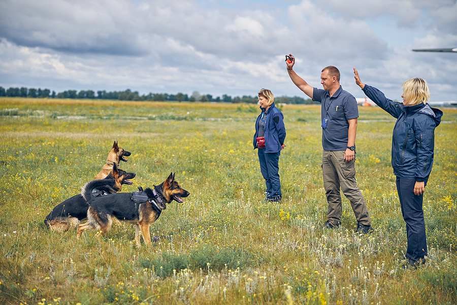
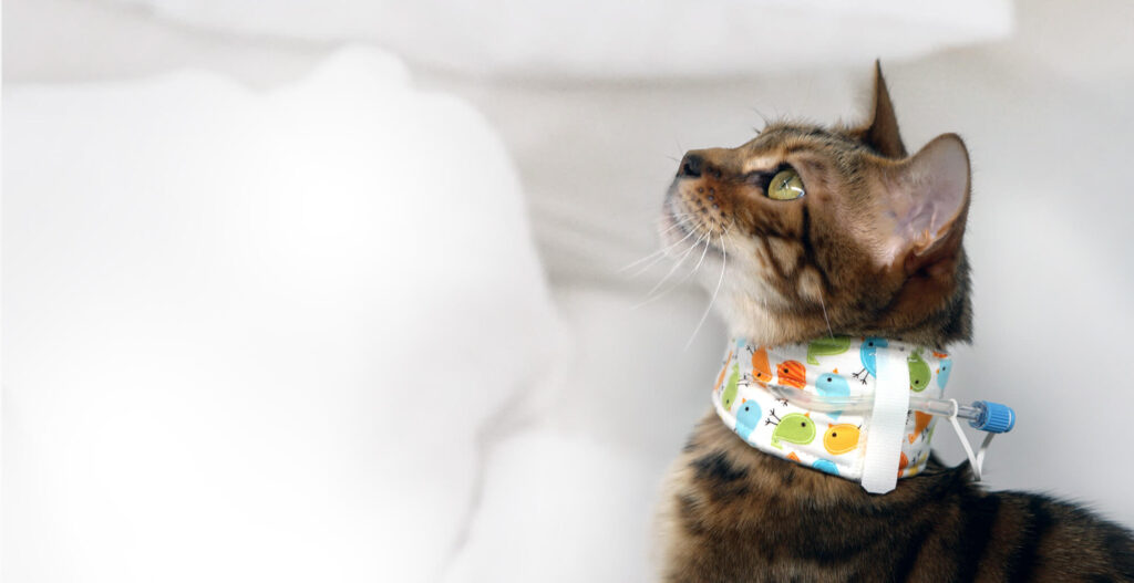
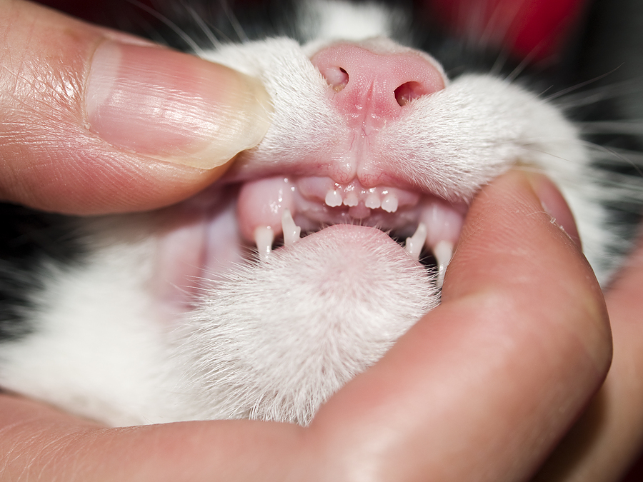

 Here’s a good rule of thumb when choosing a chew toy: if you can’t easily bend it with your hands or if you wouldn’t want to be hit in the knee with it, don’t give it to a pet. Wild dogs and wolves often have multiple fractures in their mouths due to chewing on bones.
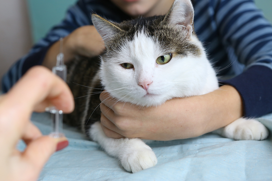
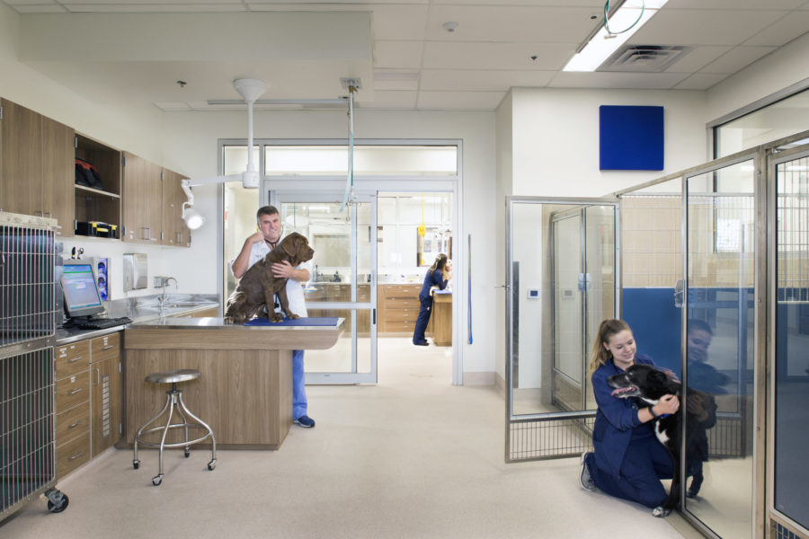
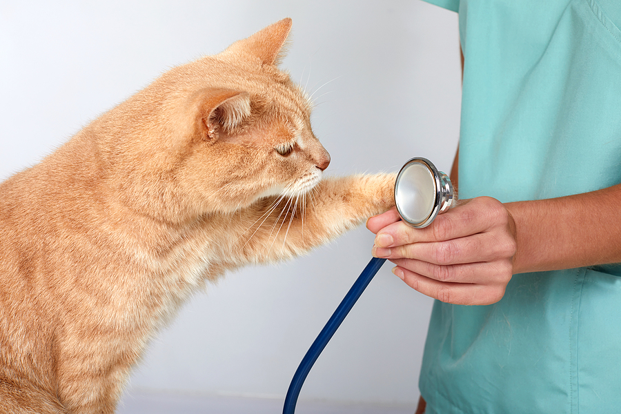
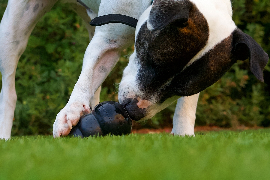
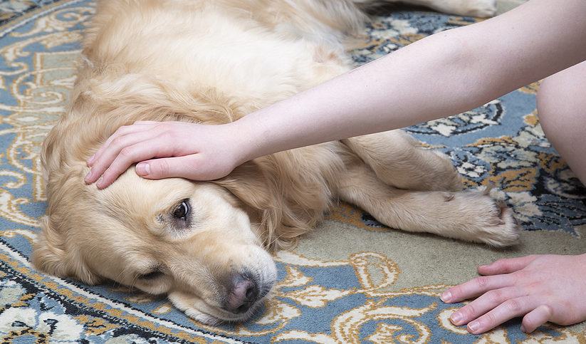
 Sponsored by our friends at Zoetis Petcare. NA-02335